One Of The Best Ways To Get People To Do Something Is To Make It Fun. A Marketing Campaign Called ‘The
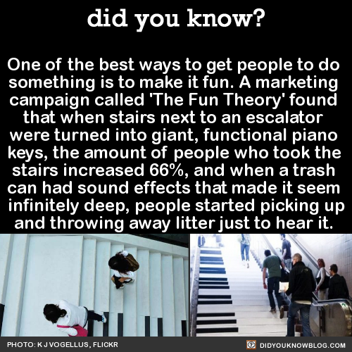
One of the best ways to get people to do something is to make it fun. A marketing campaign called ‘The Fun Theory’ found that when stairs next to an escalator were turned into giant, functional piano keys, the amount of people who took the stairs increased 66%, and when a trash can had sound effects that made it seem infinitely deep, people started picking up and throwing away litter just to hear it. Source
More Posts from Science-is-magical and Others


How Printing a 3-D Skull Helped Save a Real One
What started as a stuffy-nose and mild cold symptoms for 15-year-old Parker Turchan led to a far more serious diagnosis: a rare type of tumor in his nose and sinuses that extended through his skull near his brain.
“He had always been a healthy kid, so we never imagined he had a tumor,” says Parker’s father, Karl. “We didn’t even know you could get a tumor in the back of your nose.”
The Portage, Michigan, high school sophomore was referred to the University of Michigan’s C.S. Mott Children’s Hospital, where doctors determined the tumor extended so deep that it was beyond what regular endoscopy could see.
The team members needed to get the best representation of the tumor’s extent to ensure that their surgical approach could successfully remove the entire mass
“Parker had an uncommon, large, high-stage tumor in a very challenging area,” says Mott pediatric head and neck surgeon David Zopf, M.D. “The tumor’s location and size had me question whether a minimally invasive approach would allow us to remove the tumor completely.”
To help answer that question, teams at Mott sought an innovative approach: crafting a 3-D replica of Parker’s skull.
The model, made of polylactic acid, helped simulate the coming operation on Parker by giving U-M surgeons “an exact replica of his craniofacial anatomy and a way to essentially touch the ‘tumor’ with our hands ahead of time,” Zopf says.
Just as important, it also allowed the team to counsel Parker and his family by offering them a look at what lurked within — and, with the test run successfully complete, what would lie ahead.
A ‘pretty impressive’ model
The rare and aggressive tumor in Parker’s nose is known as juvenile nasopharyngeal angiofibroma, a mass that grows in the back of the nasal cavity and predominantly affects young male teens. Mott sees a handful of cases each year.
In Parker’s case, the tumor had two large parts: one roughly the size of an egg and the other the size of a kiwi. The mass sat right in the center of the craniofacial skeleton below the brain and next to the nerves that control eye movement and vision.
“We were obviously concerned about the risks involved in this kind of procedure, which we knew could lead to a lot of blood loss and was sensitive because it was so close to the nerves in his face,” says Karl, who praised the 3-D methodology used to aid his son. “It was pretty impressive to see the model of Parker’s skull ahead of the surgery. We had no idea this was even possible.”
Zopf, working with Erin McKean, M.D., a U-M skull base surgeon, was able to completely remove the large tumor. Kyle VanKoevering, M.D., and Sajad Arabnejad, Ph.D., aided in model preparation.
Through preoperative embolization, the blood supply to the tumor was blocked off the day before surgery to decrease blood loss. A large portion of the tumor was then detached endoscopically and removed through the mouth. The remaining mass under the brain was taken out through the nose.
Doctors took pictures of Parker’s anatomy during the surgery and, later, compared it with pictures from the model. They were nearly identical.
“Words alone can’t express how thankful we are for Parker’s talented team of surgeons at Mott,” says his mother, Heidi. “Parker is back to his old self again.”
Powerful potential
Although medical application of the technology continues to gain attention, it isn’t entirely new. Zopf and Mott teams have used 3-D printing for almost five years.
Groundbreaking 3-D printed splints made at U-M have helped save the lives of babies with severe tracheobronchomalacia, which causes the windpipe to periodically collapse and prevents normal breathing. Mott has also used 3-D printing on a fetus to plan for a potentially complicated birth.
“We are finding more and more uses for 3-D printing in medicine,” Zopf says. “It is proving to be a powerful tool that will allow for enhanced patient care.”
Based on success in patients such as Parker and continued collaboration, it’s a concept that appears poised to thrive.
“Because of the team approach we’ve established at the University of Michigan between otolaryngology and biomedical engineering, the printed models can be designed and rapidly produced at a very low cost,” Zopf says. “Michigan is one of only a few places in the nation and world that has the capacity to do this.”

From vision to hand action
Our hands are highly developed grasping organs that are in continuous use. Long before we stir our first cup of coffee in the morning, our hands have executed a multitude of grasps. Directing a pen between our thumb and index finger over a piece of paper with absolute precision appears as easy as catching a ball or operating a doorknob. The neuroscientists Stefan Schaffelhofer and Hansjörg Scherberger of the German Primate Center (DPZ) have studied how the brain controls the different grasping movements. In their research with rhesus macaques, it was found that the three brain areas AIP, F5 and M1 that are responsible for planning and executing hand movements, perform different tasks within their neural network. The AIP area is mainly responsible for processing visual features of objects, such as their size and shape. This optical information is translated into motor commands in the F5 area. The M1 area is ultimately responsible for turning this motor commands into actions. The results of the study contribute to the development of neuroprosthetics that should help paralyzed patients to regain their hand functions (eLife, 2016).
The three brain areas AIP, F5 and M1 lay in the cerebral cortex and form a neural network responsible for translating visual properties of an object into a corresponding hand movement. Until now, the details of how this “visuomotor transformation” are performed have been unclear. During the course of his PhD thesis at the German Primate Center, neuroscientist Stefan Schaffelhofer intensively studied the neural mechanisms that control grasping movements. “We wanted to find out how and where visual information about grasped objects, for example their shape or size, and motor characteristics of the hand, like the strength and type of a grip, are processed in the different grasp-related areas of the brain”, says Schaffelhofer.
For this, two rhesus macaques were trained to repeatedly grasp 50 different objects. At the same time, the activity of hundreds of nerve cells was measured with so-called microelectrode arrays. In order to compare the applied grip types with the neural signals, the monkeys wore an electromagnetic data glove that recorded all the finger and hand movements. The experimental setup was designed to individually observe the phases of the visuomotor transformation in the brain, namely the processing of visual object properties, the motion planning and execution. For this, the scientists developed a delayed grasping task. In order for the monkey to see the object, it was briefly lit before the start of the grasping movement. The subsequent movement took place in the dark with a short delay. In this way, visual and motor signals of neurons could be examined separately.
The results show that the AIP area is primarily responsible for the processing of visual object features. “The neurons mainly respond to the three-dimensional shape of different objects”, says Stefan Schaffelhofer. “Due to the different activity of the neurons, we could precisely distinguish as to whether the monkeys had seen a sphere, cube or cylinder. Even abstract object shapes could be differentiated based on the observed cell activity.”
In contrast to AIP, area F5 and M1 did not represent object geometries, but the corresponding hand configurations used to grasp the objects. The information of F5 and M1 neurons indicated a strong resemblance to the hand movements recorded with the data glove. “In our study we were able to show where and how visual properties of objects are converted into corresponding movement commands”, says Stefan Schaffelhofer. “In this process, the F5 area plays a central role in visuomotor transformation. Its neurons receive direct visual object information from AIP and can translate the signals into motor plans that are then executed in M1. Thus, area F5 has contact to both, the visual and motor part of the brain.”
Knowledge of how to control grasp movements is essential for the development of neuronal hand prosthetics. “In paraplegic patients, the connection between the brain and limbs is no longer functional. Neural interfaces can replace this functionality”, says Hansjörg Scherberger, head of the Neurobiology Laboratory at the DPZ. “They can read the motor signals in the brain and use them for prosthetic control. In order to program these interfaces properly, it is crucial to know how and where our brain controls the grasping movements”. The findings of this study will facilitate to new neuroprosthetic applications that can selectively process the areas’ individual information in order to improve their usability and accuracy.
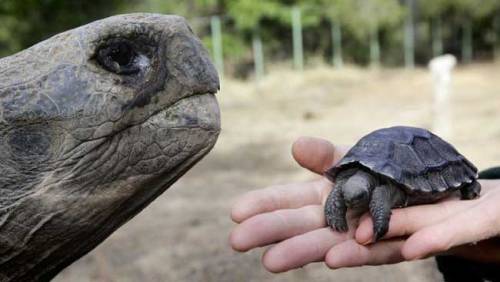
Baby Tortoises Show Up In The Galapagos For The First Time In Over A Century
There hadn’t been one single baby tortoise sighting in more than a century on the Galapagos Island of Pinzon, until a small group of the tiny, shelled youngsters were spotted this year.
The recent births are helping to pull the critically endangered animals back from the brink of extinction after they were nearly laid to waste as a result of human activity.
This is huge news for a species that has been struggling to survive for a century, relying on humans raising young tortoises bred in captivity until they are large enough to not fall prey to rats and predators.

i’m proud of them


Timelapse of Europa & Io orbiting Jupiter, shot from Cassini during its flyby of Jupiter
![Source [x]](https://64.media.tumblr.com/c67e16b83f88fe48da4656147229f385/tumblr_nz90mw4OiA1u1i4d4o1_500.jpg)
![Source [x]](https://64.media.tumblr.com/a60c74bcd55fb8555c574abb6eb4a0aa/tumblr_nz90mw4OiA1u1i4d4o2_500.jpg)
![Source [x]](https://64.media.tumblr.com/35e92a53a2e18de1283034cd6d7999ea/tumblr_nz90mw4OiA1u1i4d4o3_500.jpg)
![Source [x]](https://64.media.tumblr.com/4c1566aefd626f49bf3bfaf0c2e04616/tumblr_nz90mw4OiA1u1i4d4o4_500.jpg)
![Source [x]](https://64.media.tumblr.com/61c3673d595a1d1ddb2bd9c6d25862ae/tumblr_nz90mw4OiA1u1i4d4o5_500.jpg)
![Source [x]](https://64.media.tumblr.com/3830b9a2ede2b1021bb8596b1bc4bb6f/tumblr_nz90mw4OiA1u1i4d4o6_500.jpg)
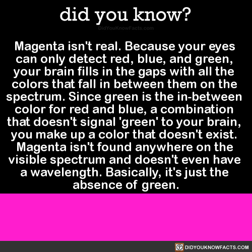
-
 blue-eyed-boss liked this · 1 year ago
 -
 bleeblu liked this · 1 year ago
 -
 comptenefekenz liked this · 1 year ago
 -
 beyondtraditional reblogged this · 1 year ago
 -
 sizzlingpizzapoetry liked this · 2 years ago
 -
 cookiesthegreat reblogged this · 3 years ago
 -
cookiesthegreat reblogged this · 3 years ago
-
 nahobme reblogged this · 3 years ago
 -
nahobme liked this · 3 years ago
-
 jamiekendal reblogged this · 3 years ago
 -
 draw-ink liked this · 3 years ago
 -
 bubblegum-bee-otch liked this · 3 years ago
 -
 oxymitch-archive reblogged this · 3 years ago
 -
 thatforestprince liked this · 3 years ago
 -
 demondark81 liked this · 4 years ago
 -
 g1ngan1nja liked this · 4 years ago
 -
 reallybadjokereally liked this · 4 years ago
 -
 pan-the-mischievous reblogged this · 4 years ago
 -
pan-the-mischievous liked this · 4 years ago
-
 toswimamongthestars reblogged this · 4 years ago
 -
 pocklepocl reblogged this · 4 years ago
 -
 pinkbat5 liked this · 4 years ago
 -
 unbearably-bear liked this · 4 years ago
 -
 nakatsoo reblogged this · 4 years ago
 -
 thelittlestprincess7 liked this · 4 years ago
 -
 mock-kett liked this · 4 years ago
 -
 selkramazed reblogged this · 4 years ago
 -
 angulface reblogged this · 4 years ago
 -
 lous-blue liked this · 4 years ago
 -
 pupuseriazag liked this · 4 years ago
 -
 senseitophat liked this · 4 years ago
 -
 popcornaddict500 liked this · 4 years ago
