WIP

WIP
More Posts from Bio100cia-blog and Others
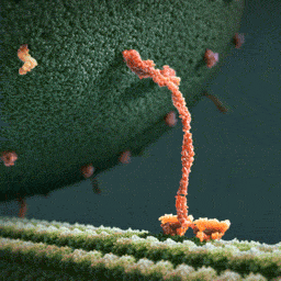
Kinesin (a motor protein) pulling some kind of vesicle along some kind of cytoskeletal filament.

Image of the Week - September 10, 2018
CIL:39062 - http://cellimagelibrary.org/images/39062
Description: This light micrograph shows the outside edge of two seminiferous tubules of a mouse testis. This section is a 1um thick transverse section, stained with toluidine blue to highlight the cells, with the nucleus staining a darker blue. The dark blue line separating the two seminiferous tubules consists mostly of myoid (muscle) tissue. The majority of cells seen in this image (arranged in layers) are germ cells, which, by repeated cell division, eventually produce spermatozoa. Chromosomes are visible in most of the nuclei of the cells. The cell with a deeply-stained nucleus and even darker nucleolus is a Sertoli cell (‘nurse cell’) that has fine cytoplasmic extensions branching between the other cells to nurture the developing germ cells.
Author: Spike Walker
Licensing: Attribution-NonCommercial-NoDerivs 2.0 UK: England & Wales (CC BY-NC-ND 2.0 UK)
![Amazing Shots Of A Cell Splitting In Two! [Photos Via María José Calasanz]](https://64.media.tumblr.com/7f3390c49f6bc9a712f44bc14f59b0f3/tumblr_omva19tRli1s04h2ho1_500.jpg)
Amazing shots of a cell splitting in two! [Photos via María José Calasanz]

The microscopic diversity of a single droplet of water.

With all the biochemistry jokes being thrown around, here’s one from my end of the biology spectrum!

Ahora todo tiene sentido xD
Clostridium difficile

Acinetobacter

Streptococcus pneumoniae

Extended-spectrum β-lactamase (ESBLs)

Enterococcus

Staphylococcus aureus

Pseudomonas aeruginosa
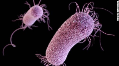
Enterobacteriaceae
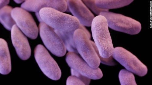
Shigella

Neisseria gonorrhoeae

Candida

Campylobacter

Tuberculosis

Salmonella


ProtoPhotosynthesis™
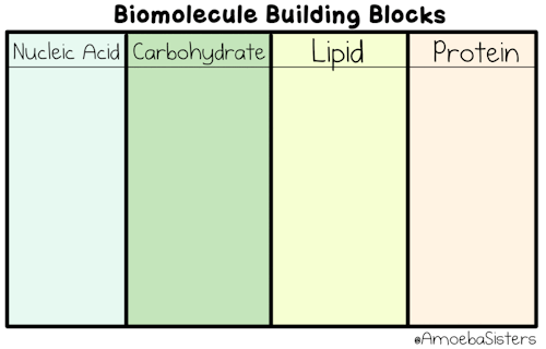
The building blocks of the four biomolecules! You can learn more about them in our video.
-
 magpie-superstition reblogged this · 5 years ago
 -
 elliestudycorner reblogged this · 5 years ago
 -
 mind-over-looks liked this · 5 years ago
 -
 kasuga707 liked this · 5 years ago
 -
 meowtasticworld reblogged this · 5 years ago
 -
 mondfamilie liked this · 5 years ago
 -
 quite-a-guy reblogged this · 5 years ago
 -
 marie-curie liked this · 5 years ago
 -
 czescihalo reblogged this · 5 years ago
 -
 rizblogs liked this · 5 years ago
 -
 el-espejo-del-tiempo liked this · 5 years ago
 -
 whoamikiding15 liked this · 5 years ago
 -
 a-certain-edge liked this · 5 years ago
 -
 corrodent reblogged this · 5 years ago
 -
corrodent liked this · 5 years ago
-
 a-little-bit-of-lots-of-stuff liked this · 6 years ago
 -
 strifetamer reblogged this · 6 years ago
 -
 godismymedicine liked this · 6 years ago
 -
 lilhunteronacase liked this · 6 years ago
 -
 atomic9uy liked this · 6 years ago
 -
 bolioptics liked this · 6 years ago
 -
 brandef reblogged this · 6 years ago
 -
 loopinthrough reblogged this · 6 years ago
 -
 bio100cia-blog reblogged this · 6 years ago
 -
 bleed-lips liked this · 6 years ago
 -
 awesome-collateral-effects reblogged this · 6 years ago
 -
 consistently-constant liked this · 6 years ago
 -
 monserrat1417-blog liked this · 6 years ago
 -
 poponchis liked this · 6 years ago
 -
 cruelfeast reblogged this · 6 years ago
 -
 la-luna-del-fisico-blog reblogged this · 6 years ago
 -
 nestakins liked this · 7 years ago
 -
 jack-o-infeliz liked this · 7 years ago
 -
 bluenebulosa liked this · 7 years ago
 -
 t-b-a-blr-blog liked this · 7 years ago
 -
 sparkleskissa liked this · 7 years ago
 -
 tennugozen liked this · 7 years ago
 -
 intisarsh liked this · 7 years ago
 -
 epicchameleon liked this · 7 years ago
